954-792-4663
 Hormonal Decline Leads to Degenerative Disease
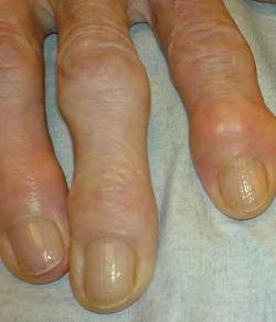
Degenerative osteoarthritis is common in post-menopausal women resulting in deformity, swelling and pain in finger joints and knee joints. Recent medical research shows that menopausal estrogen deficiency is a direct cause. In addition, bioidentical hormone replacement (including estrogen) prevents osteoarthritis.
Above left image: Typical changes of osteoarthritis of the finger joints, courtesy of wikimedia commons.
According to Dr Felson in Current Opinions in Rheumatology May 1998, osteoarthritis increases dramatically in women after menopausal age of 50 years. Medical studies following post menopausal women bio identical hormone users report reduced osteoarthritis when compared to women not using. This suggests a role for estrogen supplements for prevention of osteoarthritis in women after age 50. (15)
In 2009, Dr. Herrero- Beaumont from Spain reviewed the medical literature from 1952 to 2008 and found three causes for osteoarthritis and one of them is estrogen deficiency. (1) His article was published in Seminars in Arthritis and Rheumatism 2009.(1)
In another publication in 2009 Arthritis Research and Therapy, Dr Herrero Beaumont says:
“There is now increasing evidence that estrogens influence the activity of joint tissues through complex molecular pathways that act at multiple levels.”(2)
 A 1996 study on estrogen replacement and osteoarthritis published in the Archives of Internal Medicine from Dr Genant at the University of California, San Francisco examined 4,366 post-menopausal women over the age of 65. Hip X-Rays were used to assess osteoarthritis of the hip joint. They found that women who took oral estrogen had a 38% reduced risk osteoarthritis (OA) of the hip. Women who used Estrogen for 10 years or longer had a 43% reduction in OA of the hip. The authors concluded that:
” Postmenopausal estrogen replacement therapy may protect against OsteoArthritis (OA) of the hip.” (3)
The Framingham Study on Arthritis of the Knee was published in Arthritis and Rheum in 1998 by Dr Zhang of Boston University School of Medicine entitled, (4) They examined whether estrogen replacement therapy (ERT) prevents worsening of radiographic knee osteoarthritis (OA) in elderly women. They followed 551 post-menopausal women (over the age of 63) for 8 years with serial knee X-Rays, looking for worsening of osteoarthritis over time. The authors found a 60% decrease in osteo-arthritis in the estrogen users compared to non-users.(4)
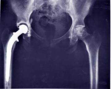
Above left image: Hip replacement could be prevented by Estrogen Replacement, image courtesy of wikimedia commons
Data from the Womens Helath Initiative Study published in 2006 showed that women receiving Premarin-alone (oral estrogen from a horse) had 12% lower rates for joint replacement for osteoarthritis.(5)
A study was in the 2010 Journal of Osteoarthritis Cartilage by Dr Riancho from Spain (6) explored the association of genetic abnormalities with severe osteoarthritis (OA) in 3147 patient who were compared to 2,381 normal controls.(6) The authors examined two abnormal genes which reduce estrogen activity. These are the gene for the Aromatase Enzyme, and the gene for the estrogen receptor (ER-Alpha), and their association with severe osteoarthritis (OA).
Women with unfavorable genotypes (a mutation) had 60% increased risk for knee arthritis. The authors conclude,
“Common genetic variations of the aromatase and ER genes are associated with the risk of severe OA of the large joints of the lower limb in a sex-specific manner. These results are consistent with the hypothesis that estrogen activity may influence the development of large-joint OA.”(6)
Thus, the genetic studies also indicate the importance of estrogen in preserving joint cartilage and preventing osteoarthritis.
Dr Tanko from Denmark summarized three decades of medical research in an article published in 2008 in Climacteric stating:
” Estrogen receptors have been identified in articular chondrocytes from various animals and humans.” …. “The effects of estrogen on articular cartilage further corroborate the due consideration of estrogen therapy for maintaining not only bone but also cartilage health in postmenopausal women.” . (8)
A study published in Arthritis Rheumatism 2002 by Dr Ham from the University of Minnesota examined estrogen replacement therapy on the severity of osteoarthritis of the knee joint in postmenopausal female monkeys (after surgical removal of the ovaries).(7)
After three years of estrogen treatment using Premarin the monkeys were sacrificed and knee joints examined under the microscope. The authors found that cartilage lesions of osteoarthritis were significantly less severe in the animals given estrogen replacement compared with those in the control group. The authors concluded,
” These results demonstrate that long-term estrogen replacement significantly reduces the severity of osteoarthritis.”(7)
A 2006 study by Tanko from Denmark published in Arthritis Rheumatism found that estrogen prevented joint and cartilage degradation in an animal model with mice. The authors found that treatment of the animals with estrogen prevents collagen deterioration with the greatest benefits for prompt initiation of hormone replacement after menopause. Estrogen protected the cartilage cells, the chondrocytes. from deterioration.. (9)
Similarly in pigs, researchers found estrogen prevents cartilage degradation. Published by Claassen from Germany in 2002 Annals of Anatomy, their study investigated how estrogen deficiency affects the articular cartilage. When they examined animals with estrogen deficiency, they found the articular cartilage underwent degradation, similar to changes of aging .(10)
A guinea pig study by Dr Dai from China was published in the Chinese medical literature in 2005 (11) The authors used the scanning electron microscope (SEM) and transmission electron microscope (TEM) to analyze cartilage degeneration in joints after ovariectomy (surgical removal of the ovaries), a form of castration which produces estrogen deficiency in the animals. The authors looked at estrogen receptors and serum levels of estrogen. They indeed found estrogen receptors (ER) in the cartilage of the guinea pigs. They also found joint cartilage degeneration detected by electron microscopy at 6 weeks, and more severe degeneration at 12 weeks (post-ovarectomy) compared to controls. The author concludes that
“Bilateral ovariectomy in the guinea pig leads to severe osteoarthritis “.(11)
A study published in 1997 in Osteoarthritis Cartilage by Dr Turner from Colorado State University looked at estrogen replacement with estradiol implants in ovarectomized sheep.(12) After twelve months, the articular cartilage from the knee joints were carefully evaluated. Ovarectomy, with its attendant estrogen deficiency had a significant deleterious effect on articular cartilage. Treatment with estradiol, a bioidentical hormone, reversed these deleterious effects, and maintained structural integrity of the joints. (12)
Another sheep study published in 2005 in Osteoarthritis Cartilage by Dr MA Cake from Australia examined the effect of estrogen depletion (ovariectomy) on articular cartilage of the joints, and the production nitric oxide synthase (iNOS). (13) At 26 weeks after removal of the ovaries, the joints were studied histologicaly, and amounts for nitric oxide studied.
In the estrogen deficient animals, cartilage thickness was reduced, along with arthritic changes and up-regulated nitric oxide production.(13) The authors conclude,
“estrogen depletion caused regional thinning of femoro-tibial cartilage, with biomechanical and histological changes suggestive of a disturbance in proteoglycan and collagen.“(13)
In 2008, Dr Sniekers of the Netherlands summarized all preceding animal studies in a report published in Osteoarthritis Cartilage. The author noted that the prevalence of osteoarthritis increases dramatically in women after the age of 50 with onset of menopausal estrogen deficiency. Animal models are useful to evaluate this. The author found 11 of 14 animal studies showed that ovarectomy (surgically induced estrogen deficiency) resulted in cartilage damage, indicating considerable evidence for a relation between cartilage degeneration and estrogen deficiency (ovarectomy) in animals. (14)
Conclusion
There is now overwhelming evidence from both animal and human studies that estrogen deficiency causes osteoarthritis, and that bioidentical estrogen replacement after menopause prevents joint degeneration.
Articles with related interest:
Arthritis and Nightshade Vegetables
The Safety of Bio-Identical Hormones
The Importance of BioIdentical Hormones
Bioidentical Hormones Prevent Arthritis
Bioidentical Hormone Estrogen Prevents Heart Disease
Morning Rounds With Steven Economou MD
Waking Up from the Synthetic Hormone Nightmare
HRT Does Not Cause Breast Cancer
Bioidentical Hormones Beneficial After Hysterectomy
Jeffrey Dach MD
7450 Griffin Road, Suite 190
Davie, Fl 33314
954-792-4663
www.jeffreydach.com
www.drdach.com
www.naturalmedicine101.com
www.bioidenticalhormones101.com
www.truemedmd.com
Click Here for: Dr Dach’s Online Store for Pure Encapsulations Supplements
Click Here for: Dr Dach’s Online Store for Nature’s Sunshine Supplements
Link to this article:http://wp.me/P3gFbV-5s
Links and References
1) www.ncbi.nlm.nih.gov/pubmed/19589561
Semin Arthritis Rheum. 2009 Oct;39(2):71-80. Primary osteoarthritis no longer primary: three subsets with distinct etiological, clinical, and therapeutic characteristics. Herrero-Beaumont G, Roman-Blas JA, Castañeda S, Jimenez SA. Source Bone and Joint Research Unit, Service of Rheumatology, Fundación Jiménez Díaz, Universidad Autónoma, Madrid, Spain.
Osteoarthritis (OA) has been historically divided into primary and secondary. Primary OA has been defined as an idiopathic condition developing in previously undamaged joints in the absence of an obvious causative mechanism. During the last few years a large amount of evidence has provided new insights into the biochemistry and molecular biology of cartilage, subchondral bone, and other articular tissues, which suggest distinct etiopathogenetic mechanisms in some forms of primary OA. OBJECTIVE: To propose an etiopathogenic classification of primary OA in the light of the significant progress in the understanding of the disease.
METHODS: A review of the literature was performed by searching the Medline and PubMed databases from 1952 to November 2008 using the following keywords: genetic alteration, heritability, estrogen, menopause, and aging either alone or in various combinations with joint, cartilage, subchondral bone, synovium, ligaments, muscle, tendons, OA, and osteoporosis.
RESULTS: Numerous studies have shown that genetic alterations, menopause-related estrogen deficiency, and aging play crucial roles in the molecular pathophysiological events involved in the process of cartilage and joint damage and thus in development of OA.
We propose classifying primary OA into 3 distinct although interrelated subsets:
type I OA, genetically determined;
type II OA, estrogen hormone dependent;
and type III OA, aging related.
CONCLUSIONS: The 3 proposed subsets of OA display distinct etiological, clinical, and therapeutic characteristics and should therefore no longer be considered to be “Primary OA.”
2) www.ncbi.nlm.nih.gov/pmc/articles/PMC2787275/?tool=pubmed
FULL TEXT – Arthritis Res Ther. 2009; 11(5): 241.
Osteoarthritis associated with estrogen deficiency
Jorge A Roman-Blas,1,2 Santos Castañeda,3 Raquel Largo,1 and Gabriel Herrero-Beaumontcorresponding author1 1Bone and Joint Research Unit, Service of Rheumatology, Fundación Jiménez Díaz, Universidad Autónoma, Madrid 28040, Spain
Osteoarthritis (OA) affects all articular tissues and finally leads to joint failure. Although articular tissues have long been considered unresponsive to estrogens or their deficiency, there is now increasing evidence that estrogens influence the activity of joint tissues through complex molecular pathways that act at multiple levels.
3) www.ncbi.nlm.nih.gov/pubmed/8862099
Arch Intern Med. 1996 Oct 14;156(18):2073-80.
Association of estrogen replacement therapy with the risk of osteoarthritis of the hip in elderly white women.
Study of Osteoporotic Fractures Research Group. Nevitt MC, Cummings SR, Lane NE, Hochberg MC, Scott JC, Pressman AR, Genant HK, Cauley JA. Source Department of Epidemiology and Biostatistics, University of California, San Francisco, USA.
Abstract OBJECTIVE: To determine whether postmenopausal estrogen replacement therapy is associated with a reduced risk of radiographic findings of osteoarthritis (OA) of the hip. DESIGN: Cross-sectional study.
SUBJECTS: White women (N = 4366; age, > or = 65 years) who were participants in a cohort study of osteoporotic fractures.
MEASUREMENTS AND METHODS: Radiographs of the pelvis that were obtained in all subjects were assessed for radiographic features of OA of the hip on a summary scale of 0 (none) to 4 (severe OA). Postmenopausal estrogen use was assessed by interview. The association of current and past oral estrogen use with OA of the hip was analyzed by using logistic regression, adjusting for potential confounding variables (eg, indicators of osteoporosis and correlates of estrogen use).
RESULTS: Five hundred thirty-nine women (12.3%) had mild or greater radiographic findings of OA of the hip in at least 1 hip, and 214 women (4.9%) had moderate to severe findings; 17% and 24% of the women were current and past users of oral estrogen, respectively. Women who were currently using oral estrogen had a significantly reduced risk of any OA of the hip (adjusted odds ratio [OR], 0.62; 95% confidence interval [CI], 0.49-0.86) and moderate to severe manifestation of disease (OR, 0.54; 95% CI, 0.33-0.88). Current users who had taken estrogen for 10 years or longer had a greater reduction in the risk of any OA of the hip (OR, 0.57; 95% CI, 0.40-0.82) compared with that of users for less than 10 years (OR, 0.75; 95% CI, 0.47-1.24). Current estrogen use for 10 years or longer was associated with a nonsignificant trend for a reduced risk of moderate to severe symptomatic disease (OR, 0.59; 95% CI, 0.28-1.29).
CONCLUSION: Postmenopausal estrogen replacement therapy may protect against OA of the hip in elderly white women.
4) www.ncbi.nlm.nih.gov/pubmed/9778229
Arthritis Rheum. 1998 Oct;41(10):1867-73.
Estrogen replacement therapy and worsening of radiographic knee osteoarthritis: the Framingham Study.
Zhang Y, McAlindon TE, Hannan MT, Chaisson CE, Klein R, Wilson PW, Felson DT. Source Boston University School of Medicine, Massachusetts, USA.
Abstract OBJECTIVE: To examine whether estrogen replacement therapy (ERT) prevents worsening of radiographic knee osteoarthritis (OA) in elderly women. METHODS: A total of 551 women ages 63-91 years (mean age 71) in the Framingham Study were followed up from biennial examination 18 (1983-1985) to examination 22 (1992-1993). Data on postmenopausal ERT were obtained every 2 years. Subjects were classified into 3 groups according to their estrogen use at biennial examination 18: never users (n = 349), past users (n = 162), and current users (n = 40). Women received anteroposterior weight-bearing knee radiographs at examinations 18 and 22. Using the Kellgren and Lawrence criteria, global radiographic knee OA was assessed, (grade range 0-4) and individual radiographic features, such as osteophytes and joint space narrowing, were scored from 0 to 3. Worsening was defined as either development of radiographic OA that was not present at baseline (incident OA) or progression of baseline radiographic OA by > or =1 Kellgren and Lawrence grade (progressive OA). Potential confounding factors included age, body mass index, weight change, smoking, knee injury, physical activity level, and bone mineral density at the femoral neck. RESULTS: During 8 years of followup, 17.4% of knee radiographic scores worsened by 1 grade and 5.8% by 2 or 3 grades among never users of ERT.
Among current estrogen users, only 11.7% of knee radiographic scores worsened by 1 grade and none worsened by more than 1 grade. After adjusting for age and other potential confounding factors, the relative risk of incident radiographic knee OA in comparison with never users of estrogen was 0.8 (95% confidence interval [95% CI] 0.5-1.4) in past users and 0.4 (95% CI 0.1-3.0) in current users. Current use of estrogen also showed a trend toward decreased risk of progressive knee OA compared with never use (odds ratio [OR] 0.5, 95% CI 0.1-2.9). When both incident and progressive radiographic knee OA cases were combined, current ERT use had a 60% decreased risk compared with never use (OR 0.4, 95% CI 0.1-1.5).
5) www.ncbi.nlm.nih.gov/pubmed/17009251
Effect of hormone therapy on risk of hip and knee joint replacement in the Women’s Health Initiative. Cirillo DJ, Wallace RB, Wu L, Yood RA. Arthritis Rheum. 2006 Oct;54(10):3194-204. University of Iowa College of Public Health, Iowa City, IA 52242, USA.
To determine the effect of hormone therapy on arthroplasty rates.
METHODS: We examined data from the Women’s Health Initiative placebo-controlled, double-blind, randomized trials. Community-dwelling women ages 50-79 years were enrolled at 40 US clinics. Women with prior arthroplasty were excluded, yielding a sample size of 26,321 subjects. Women who had had hysterectomies (n = 10,272) were randomly assigned to receive 0.625 mg/day conjugated equine estrogens (n = 5,076), or placebo (n = 5,196), with a mean followup of 7.1 years. Those who had not had hysterectomies (n = 16,049) were randomly assigned to receive estrogen plus progestin (n = 8,240), given as 0.625 mg/day conjugated equine estrogens plus 2.5 mg/day medroxyprogesterone acetate, or placebo (n = 7,809), with a mean followup of 5.6 years. Participants reported hospitalizations, and arthroplasties were identified by procedure codes. Arthroplasties due to hip fracture were censored. Cox proportional hazards regression was used to assess hazard ratios (HRs) and 95% confidence intervals (95% CIs) using intent-to-treat methods and outcome of time to first procedure.
RESULTS: In the estrogen-alone trial, women receiving hormone therapy had significantly lower rates of any arthroplasty (HR 0.84 [95% CI 0.70- 1.00], P = 0.05).
CONCLUSION: These data suggest that hormone therapy may influence joint health, but this observed decrease in risk may be limited to unopposed estrogen and may possibly be more important in hip than in knee osteoarthritis.
GENETIC CAUSES OF Osteoarthritis
6) www.ncbi.nlm.nih.gov/pubmed/20417295
Osteoarthritis Cartilage. 2010 Jul;18(7):927-33.
Common variations in estrogen-related genes are associated with severe large-joint osteoarthritis: a multicenter genetic and functional study. Riancho JA, García-Ibarbia C, Gravani A, Raine EV, Rodríguez-Fontenla C, Soto-Hermida A, Rego-Perez I, Dodd AW, Gómez-Reino JJ, Zarrabeitia MT, Garcés CM, Carr A, Blanco F, González A, Loughlin J. Source Servicio de Medicina Interna, Hospital UM Valdecilla-IFIMAV, Universidad de Cantabria, RETICEF, Santander, Spain. rianchoj@unican.es Abstract
OBJECTIVE: Several lines of evidence suggest that estrogens influence the development of osteoarthritis (OA). The aim of this study was to explore the association of two common polymorphisms within the aromatase (CYP19A1) and estrogen receptor (ER) alpha (ESR1) genes with severe OA of the lower limbs.
METHODS: The rs1062033 (CYP19A1) and rs2234693 (ESR1) single nucleotide polymorphisms were genotyped in 5528 individuals (3147 patients with severe hip or knee OA, and 2381 controls) from four centres in Spain and the United Kingdom. Gene expression was measured in femoral bone samples from a group of patients.
RESULTS: In the global analysis, both polymorphisms were associated with OA, but there was a significant sex interaction. The GG genotype at rs1062033 was associated with an increased risk of knee OA in women [odds ratio (OR) 1.23; P=0.04]. The CC genotype at rs2234693 tended to be associated with reduced OA risk in women (OR 0.76, P=0.028, for knee OA; OR=0.84, P=0.076 for hip OA), but with increased risk of hip OA in men (OR 1.28; P=0.029). Women with unfavourable genotypes at both loci had an OR of 1.61 for knee OA (P=0.006). The rs1062033 genotype associated with higher OA risk was also associated with reduced expression of the aromatase gene in bone.
CONCLUSIONS: Common genetic variations of the aromatase and ER genes are associated with the risk of severe OA of the large joints of the lower limb in a sex-specific manner. These results are consistent with the hypothesis that estrogen activity may influence the development of large-joint OA.
Estrogen Replacement in Ovarectomized Monkeys Prevents OA
7) www.ncbi.nlm.nih.gov/pubmed/12124881
Arthritis Rheum. 2002 Jul;46(7):1956-64.
Effects of long-term estrogen replacement therapy on osteoarthritis severity in cynomolgus monkeys. Ham KD, Loeser RF, Lindgren BR, Carlson CS. Source Dpartment of Veterinary Pathology, College of Veterinary Medicine, University of Minnesota, St. Paul, 55108, USA. hamx0012@tc.umn.edu Abstract
OBJECTIVE: To determine the effects of long-term estrogen replacement therapy (ERT) on the severity of osteoarthritis (OA) of the knee joint in surgically postmenopausal (bilaterally ovariectomized) female monkeys. A secondary aim was to evaluate the effect of soy phytoestrogen (SPE) treatment on the severity of OA.
METHODS: Feral adult female cynomolgus macaques were ovariectomized bilaterally and then randomly divided into 3 age- and weight-matched treatment groups. For 3 years, the first group received ERT with conjugated equine estrogens, the second group received SPE, and the third group received no treatment (controls). At necropsy, histologic lesions of OA were graded, and the area and thickness of cartilage and subchondral bone were measured. The data were summarized by principal components analysis, and the resulting factors and individual variables were compared using analysis of variance and analysis of covariance (age and weight as covariates).
RESULTS: Cartilage lesions of OA were significantly less severe in the animals given ERT compared with those in the control group. This treatment effect remained significant when adjusted for age and weight. The factor representing subchondral bone was significantly higher, but the number of osteophytes was lower, in the ERT group compared with the control group. SPE treatment had no significant effect on cartilage or bone lesions of OA.
CONCLUSION: These results demonstrate that long-term ERT significantly reduces the severity of OA lesions in this animal model.
8) www.ncbi.nlm.nih.gov/pubmed/18202960
Climacteric. 2008 Feb;11(1):4-16.
An update review of cellular mechanisms conferring the indirect and direct effects of estrogen on articular cartilage. Tankó LB, Søndergaard BC, Oestergaard S, Karsdal MA, Christiansen C. Source Nordic Bioscience, Herlev, Denmark. Abstract
OBJECTIVE: To review cellular mechanisms that have been proposed to mediate the indirect and direct effects of estrogen on articular cartilage, and to outline the remaining clinical questions that need to be clarified before utilizing the beneficial effects of estrogen for the prevention of osteoarthritis in early postmenopausal women.
DESIGN: Summary of original research papers and reviews listed in Pubmed (1980-2007).
RESULTS: Estrogen receptors have been identified in articular chondrocytes from various animals and humans. Molecular studies showed that estrogen can elicit genomic and rapid non-genomic effects on various cell types, including chondrocytes, and the latter effects are only inducible in females. In addition to direct effects, estrogen can also affect the homeostasis of articular cartilage by modulating the expression/production of different molecules such as various growth factors, inflammatory cytokines, matrix metalloproteinases, and reactive oxygen species. Moreover, in vivo observation argues for the notion that inhibition of subchondral bone turnover is also part of the mechanisms by which estrogen (and antiresorptive agents in general) can protect against joint degradation. Published studies undertaken at cellular, tissue, and in vivo levels illustrate that the effect of estrogen on cartilage may depend on the dose applied, the administration route, the time of initiation, and whether it is combined with a progestin.
CONCLUSIONS: The herein reviewed direct and indirect effects of estrogen on articular cartilage further corroborate the due consideration of estrogen therapy for maintaining not only bone but also cartilage health in postmenopausal women. Future studies in postmenopausal women are needed to clarify whether the efficacy of estrogen therapy can be further optimized by using other forms of estrogen, other progestins, or by initiating the therapy in the peri- or early postmenopausal period.
Animal Studies
9) www.ncbi.nlm.nih.gov/pubmed/16871544
Arthritis Rheum. 2006 Aug;54(8):2441-51.
Effects of ovariectomy and estrogen therapy on type II collagen degradation and structural integrity of articular cartilage in rats: implications of the time of initiation. Oestergaard S, Sondergaard BC, Hoegh-Andersen P, Henriksen K, Qvist P, Christiansen C, Tankó LB, Karsdal MA. Source Nordic Bioscience, Herlev, Denmark.
Abstract OBJECTIVE: To investigate how the time of initiation influences the effects of estrogen therapy on type II collagen (CII) turnover and the structural integrity of articular cartilage in ovariectomized rats and to determine whether estrogen exerts direct effects on the catabolic function of chondrocytes ex vivo.
METHODS: A total of 46 Sprague-Dawley rats were distributed into 1 of the following treatment groups:
1) ovariectomy,
2) ovariectomy plus early estrogen therapy,
3) ovariectomy plus delayed estrogen therapy, or
4) sham operation.
Cartilage turnover was estimated by measuring the serum levels of C-telopeptide of type II collagen (CTX-II). Cartilage lesions at week 9 were quantified using a published scoring technique. The presence of the CTX-II epitope in articular cartilage was assessed by immunohistochemistry. The effects of estrogen (1 -100 nM) on chondrocytes were investigated in bovine cartilage explants subjected to catabolic cytokines (tumor necrosis factor alpha [TNFalpha] and oncostatin M [OSM]).
RESULTS: In ovariectomized rats, estrogen therapy evoked significant decreases in serum CTX-II independently of the time of initiation; yet, delayed initiation resulted in diminished efficacy in terms of preventing cartilage lesions. CTX-II fragments were present in articular cartilage, colocalizing with early lesions at the cartilage surface. In untreated animals, the early relative increases in serum CTX-II were proportional to the severity of cartilage lesions at week 9 (r = 0.73, P < 0.01). Estrogen significantly and dose- dependently countered CTX-II release from TNFalpha plus OSM-stimulated cartilage explants ex vivo.
CONCLUSION: Our results suggest that estrogen counters the acceleration of CII degradation and related structural alterations, and these benefits can be maximized by early initiation after menopause. The protective effect of estrogen seems to involve direct inhibition of the catabolic function of chondrocytes.
10) www.ncbi.nlm.nih.gov/pubmed/11936193
Ann Anat. 2002 Mar;184(2):141-8.
The effect of estrogens and dietary calcium deficiency on the extracellular matrix of articular cartilage in Göttingen miniature pigs. Claassen H, Hornberger F, Scholz-Ahrens K, Schünke M, Schrezenmeir J, Kurz B. Source Anatomisches Institut der Universität Kiel, Germany.
Abstract Clinical observations have suggested that estrogens are involved in the pathogenesis of postmenopausal osteoarthritis (OA). However, positive and negative associations between the incidence of OA and serum estrogen concentrations have been reported.
In contrast to this, osteoporosis is regarded as a disease with a strong estrogen-dependent component. Moreover, there is an interaction between estrogen and calcium deficiency: calcium supplementation potentiates the effect of estrogen therapy. The present study was designed to investigate how estrogen deficiency affects the articular cartilage depending on calcium supply. The distribution of different types of glycosaminoglycans and collagens can be used as an indicator for extracellular matrix changes induced by estrogen deficiency. Different levels of dietary calcium were therefore fed to intact and ovariectomized Göttingen miniature pigs for one year before articular cartilage was harvested. The histochemical staining for heavy sulfated glycosaminoglycans in the extracellular matrix of ovariectomized miniature pigs, especially of those fed with a low calcium diet, was stronger in comparison to intact animals. In intact animals type II-collagen was immunodetected in all zones of unmineralized and mineralized articular cartilage, while immunostaining for this protein was negative to weak in the deep radiated fiber zone of ovariectomized minipigs.
These results suggest that the synthesis of heavy sulfated glycosaminoglycans and immunohistochemically detectable type II-collagen is possibly influenced by estrogen
deficiency.
In conclusion, under estrogen deficiency, the extracellular matrix of articular cartilage underwent similar changes to those observed in physiologically aging cartilage where keratan sulfate is increased as a heavy sulfated glycosaminoglycan.
11) www.ncbi.nlm.nih.gov/pubmed/16696325
J Huazhong Univ Sci Technolog Med Sci. 2005;25(6):683-6.
The relationship of the expression of estrogen receptor in cartilage cell and osteoarthritis induced by bilateral ovariectomy in guinea pig. Dai G, Li J, Liu X, Liu Q, Liu C. Source Department of Orthopaedics, Oilu Hospital Affiliated Shan Dong University, Jinan 250012, China.
Abstract To investigate the estrogen receptor (ER) expression in cartilage cell in the development of osteoarthritis induced by bilateral ovariectomy in guinea pig and to find their relationship. 30 two-month-old female guinea pigs were randomly divided into two groups (n = 15 each): sham operation (control) group and ovariectomized group (OVX);
Scanning electron microscope (SEM) and transmission electron microscope (TEM) were obtained to analysis the cartilage degeneration of the hind limb knee joint after 6 and 12 weeks of ovariectomy. Dextran-Coated-Charcoal (DCC) was taken to quantitively detect the expression of ER. The serum levels of estrogen and gestone were detected by immune contest assay.
The results showed that ER do exist in the cartilages of the guinea pigs, with higher expression in the control group than in OVX group at the same time point (P < 0.05). It was increased also at 12 th week after operation than that of preoperation. The blood serum levels of estrogen and gestone showed a similar tendency to the expression of ER. Joint cartilage degeneration detected by SEM and TEM could be found at 6 th week, but severe degenerative lesions at 12 th week in the OVX group compared with the control group (P < 0.01).
The data suggested that bilateral ovariectomy in guinea pig lead to severe osteoarthritis which might be related to the lower serum level of estrogen and the downregulation of the expression of ER in the cartilage also.
12) www.ncbi.nlm.nih.gov/pubmed/9010879
Osteoarthritis Cartilage. 1997 Jan;5(1):63-9.
Biochemical effects of estrogen on articular cartilage in ovariectomized sheep.
Turner AS, Athanasiou KA, Zhu CF, Alvis MR, Bryant HU. Source Department of Clinical Sciences, Colorado State University, Ft. Collins 80523, USA.
Abstract Cartilage is a sex-hormone-sensitive tissue but the role of estrogen in the pathogenesis of osteoarthritis (OA) remains controversial. In this study, intrinsic material properties and thickness of articular cartilage of the knee joint of ovariectomized (OVX) and estrogen-treated sheep were measured. Skeletally mature ewes (N = 36, same breed, same housing 4-5 years old) were divided into; sham treated (n = 9), OVX (N = 13), OVX plus one estradiol implant (OVXE; N = 10) and OVX plus two estradiol implants (OVX2E; N = 4).
Twelve months following sham procedure or OVX, sheep were euthanized and articular cartilage from a total of 216 points in the left femorotibial (knee) joints was tested for aggregate modulus, Poisson’s ratio, permeability, thickness and shear modulus (six sites per sheep). When all of the sites in each knee were grouped together, OVX had a significant effect on articular cartilage. The sham cartilage of all sites grouped together had a larger aggregate modulus (P = 0.001) and a larger shear modulus (P = 0.054) than the OVX tissue. No statistically significant differences were seen for permeability and thickness between OVX, sham, OVXE and OVX2E. Differences existed in biomechanical properties at the different sites that were tested. Overall, no one location tended to be lowest or highest for all variables. This biomechanical study suggests that OVX may have a detrimental effect on the intrinsic material properties of the articular cartilage of the knee, even though the cartilage of the OVX animals appeared normal.
Treatment with estradiol implants ameliorated these deleterious effects and may have helped maintain the tissue’s structural integrity. Our study supports epidemiological studies of OA in women after menopause. The protective effect of estrogen and it’s therapeutic effect remain to be further defined. This model may allow the relationship of estrogen and estrogen antagonists to be studied in greater detail, and may be valuable for the study of the pathogenesis and therapies of OA of postmenopausal women, particularly in its early stages.
13) www.ncbi.nlm.nih.gov/pubmed/16154775
Osteoarthritis Cartilage. 2005 Dec;13(12):1066-75. Epub 2005 Sep 9.
Ovariectomy alters the structural and biomechanical properties of ovine femoro-tibial articular cartilage and increases cartilage iNOS. Cake MA, Appleyard RC, Read RA, Smith MM, Murrell GA, Ghosh P. Source School of Veterinary and Biomedical Sciences, Murdoch University, WA, Australia.
Abstract OBJECTIVE: To examine the effect of oestrogen depletion produced by surgical ovariectomy on the structural and biomechanical properties of ovine femoro-tibial articular cartilage (AC), and the production of inducible nitric oxide synthase (iNOS) and nitrotyrosine by these tissues. METHODS: Six aged ewes were surgically ovariectomised (OVX), while six were used as unoperated controls. Dynamic biomechanical indentation testing of tibial plateau AC was performed at 26 weeks post-op. Histological sections of medial tibial plateau and lateral tibial plateau (LTP), medial and lateral femoral condyles (MFC, LFC) and patellar AC were examined for histopathology, toluidine blue staining intensity, and patterns of collagen birefringence intensity. Immunoreactivity for iNOS and nitrotyrosine was assessed in full-thickness biopsy plugs of LFC and patellar AC, and patellar AC explants were cultured to determine in vitro NO release.
RESULTS: Phase lag was reduced overall in LTP-AC of OVX sheep (10.9+/-2.2 degrees vs 12.1+/-2.3 degrees ; P<0.0001). Cartilage thickness was reduced in the LTP of OVX sheep (P=0.0002), in association with localised changes in dynamic shear modulus. Toluidine blue staining intensity was reduced in the patella, LFC, and MFC. Histological examination revealed greater histopathology scores in the MFC of OVX animals, and altered collagen birefringence intensity plots in the LTP. Immunostaining for iNOS was increased in patella AC (P=0.008), whilst nitrotyrosine immunoreactivity was increased in patella (P=0.03) and LFC (P<0.0001) AC. NO release by patellar AC explants was also elevated.
CONCLUSIONS: Oestrogen depletion induced by OVX caused regional thinning of femoro-tibial cartilage, with biomechanical and histological changes suggestive of a disturbance in the content and/or structural organisation of the proteoglycan and collagen macromolecular assembly. The observed up-regulation of cartilage iNOS suggests a possible mechanism for these matrix changes.
ANimal Model Summary
14) www.ncbi.nlm.nih.gov/pubmed/18280756
Osteoarthritis Cartilage. 2008 May;16(5):533-41. Epub 2008 Feb 15.
Animal models for osteoarthritis: the effect of ovariectomy and estrogen treatment – a systematic approach. Sniekers YH, Weinans H, Bierma-Zeinstra SM, van Leeuwen JP, van Osch GJ. Source Department of Orthopaedics, Erasmus MC, University Medical Center, Rotterdam, The Netherlands. Abstract
OBJECTIVE: The prevalence of osteoarthritis (OA) increases dramatically in women after the age of 50. Animal models are used to study the effects of hormone depletion [by ovariectomy (OVX)] and estrogen treatment on OA. This review summarizes these animal studies, in order to get a better insight in the role of hormones on OA.
METHOD: The literature was systematically reviewed until May 2007. The results were divided into two parts: the effect of OVX on cartilage, and the effect of estrogen treatment on cartilage. Only studies with an appropriate control group (e.g., sham-operated) were included.
RESULTS AND DISCUSSION: Eleven out of 16 animal studies showed that OVX resulted in cartilage damage. When only studies using sexually mature animals were included, we saw that 11 out of 14 studies showed a detrimental effect, indicating considerable evidence for a relation between cartilage degeneration and OVX in mature animals. The effect of estrogen treatment was inconclusive with only 11 out of 22 animal studies reporting a beneficial effect on cartilage, whereas all six studies administering selective estrogen receptor modulators (SERMs) after OVX described protective effects. The discrepancy between the studies may be caused by the large variation in experimental set-up. We suggested a list of quality criteria for animal models since standardisation of design and outcome parameters of animal experiments may help to compare different studies and to gain better insight in the role of hormones in the osteoarthritic process.
HUMAN
15) The effects of estrogen on osteoarthritis
Curr Opin Rheumatol. 1998 May;10(3):269-72. The effects of estrogen on osteoarthritis. Felson DT, Nevitt MC. Source Boston University School of Medicine, Multipurpose Arthritis and Musculoskeletal Diseases Center, MA 02118-2526, USA.
Abstract The prevalence of osteoarthritis is higher in women than men, and in women it increases dramatically in the years after menopause. These observations and others reporting a painful form of hand osteoarthritis after the menopause suggest that loss of estrogen at the time of menopause increases a woman’s risk of getting osteoarthritis. This article reviews biologic evidence for hormone sensitivity of cartilage, animal studies testing the effect of estrogen on the joints of ovariectomized animals, and human epidemiologic and clinical studies evaluating endogenous estrogen levels and estrogen replacement therapy and their relation to the occurrence of osteoarthritis. Overall the evidence for a role of estrogen in osteoarthritis is conflicting. Epidemiologic studies of women who take estrogen replacement therapy, however, consistently report that these women have a lower prevalence of osteoarthritis than women not taking estrogen, suggesting a possible therapeutic role for estrogen in osteoarthritis.
PEMF – Pulsed Electro MAgnetic Fields
www.ncbi.nlm.nih.gov/pubmed/16023006
J Orthop Res. 2005 Jul;23(4):899-908. Epub 2005 Mar 17.
Pulsed electromagnetic fields reduce knee osteoarthritic lesion progression in the aged Dunkin Hartley guinea pig. Fini M, Giavaresi G, Torricelli P, Cavani F, Setti S, Canè V, Giardino R. Source Department of Experimental Surgery, Codivilla-Putti Research Institute, Rizzoli Institute of Orthopaedics, Via di Barbiano, 1/10, 40136 Bologna, Italy.
Abstract An experimental in vivo study was performed to test if the effect of Pulsed Electromagnetic Fields (PEMFs) on chondrocyte metabolism and adenosine A2a agonist activity could have a chondroprotective effect on the knee of Dunkin Hartley guinea-pigs of 12 months with spontaneously developed osteoarthritis (OA).
After a pilot study, 10 animals were randomly divided into two groups: PEMF-treated group (6 h/day for 3 months) and Sham-treated group. Microradiography and histomorphometry were performed on the entire articular surface of knee joints used in evaluating chondropathy severity, cartilage thickness (CT), cartilage surface Fibrillation Index (FI), subchondral bone plate thickness (SBT) and histomorphometric characteristics of trabecular epiphyseal bone. The PEMF-treated animals showed a significant reduction of chondropathy progression in all knee examined areas (p<0.05). CT was significantly higher (p<0.001) in the medial tibia plateaus of the PEMF-treated group when compared to the Sham-treated group. The highest value of FI was observed in the medial tibia plateau of the Sham-treated group (p<0.05). Significant lower values were observed in SBT of PEMF-treated group in comparison to Sham-treated group in all knee examined areas (p<0.05). The present study results show that PEMFs preserve the morphology of articular cartilage and slower the progression of OA lesions in the knee of aged osteoarthritic guinea pigs. The chondroprotective effect of PEMFs was demonstrated not only in the medial tibial plateau but also on the entire articular surface of the knee.
www.ncbi.nlm.nih.gov/pubmed/17459652
Biomed Pharmacother. 2008 Dec;62(10):709-15. Epub 2007 Apr 3.
Effect of pulsed electromagnetic field stimulation on knee cartilage, subchondral and epyphiseal trabecular bone of aged Dunkin Hartley guinea pigs.
Fini M, Torricelli P, Giavaresi G, Aldini NN, Cavani F, Setti S, Nicolini A, Carpi A, Giardino R. Source Laboratory of Experimental Surgery, Research Institute Codivilla-Putti, Rizzoli Orthopaedic Institute, Bologna, Italy.
Abstract It has been demonstrated that pulsed electromagnetic field (PEMF) stimulation has a chondroprotective effect on osteoarthritis (OA) progression in the knee joints of the 12-month-old guinea pigs. The aim of the present study was to discover whether the therapeutic efficacy of PEMFs was maintained in older animals also in more severe OA lesions. PEMFs were administered daily (6 h/day for 6 months) to 15-month-old guinea pigs. The knee joints (medial and lateral tibial plateaus, medial and lateral femoral condyles) were evaluated by means of a histological/histochemical Mankin modified by Carlsson grading score and histomorphometric measurements of cartilage thickness (CT), fibrillation index (FI), subchondral bone thickness (SBT) and epiphyseal bone microarchitecture (bone volume: BV/TV; trabecular thickness: Tb.Th; trabecular number: Tb.N; trabecular separation: Tb.SP). Periarticular knee bone was also evaluated with dual X-ray absorptiometry (DXA). PEMF stimulation significantly changed the progression of OA lesions in all examined knee areas. In the most affected area of the knee joint (medial tibial plateau), significant lower histochemical score (p<0.0005), FI (p<0.005), SBT (p<0.05), BV/TV (p<0.0005), Tb.Th (p<0.05) and Tb.N (p<0.05) were observed while CT (p<0.05) and Tb.Sp (p<0.0005) were significantly higher than in SHAM-treated animals. DXA confirmed the significantly higher bone density in SHAM-treated animals.
Even in the presence of severe OA lesions PEMFs maintained a significant efficacy in reducing lesion progression.
Low Level Laser for OA
www.integralnatmed.com/pdf/LLLT_Knee_Osteoarthritis_Photomedicine_2009.pdf
Photomedicine and Laser Surgery Volume 27, Number 4, 2009
The Effect of Low-Level Laser in Knee Osteoarthritis: A Double-Blind, Randomized, Placebo-Controlled Trial Be´ la Heged}us, M.D.,1 La´ szlo´ Viharos, Ph.D.,2 Miha´ ly Gervain, Ph.D.,3 and Ma´ rta Ga´ lfi, Ph.D.4 Introduction:
Low-level laser therapy (LLLT) is thought to have an analgesic effect as well as a biomodulatory effect on microcirculation. This study was designed to examine the pain-relieving effect of LLLT and possible microcirculatory changes measured by thermography in patients with knee osteoarthritis (KOA). Materials and Methods: Patients with mild or moderate KOA were randomized to receive either LLLT or placebo LLLT. Treatments were delivered twice a week over a period of 4wk with a diode laser (wavelength 830 nm, continuous wave, power 50mW) in skin contact at a dose of 6 J=point. The placebo control group was treated with an ineffective probe (power 0.5mW) of the same appearance. Before examinations and immediately, 2 wk, and 2 mo after completing the therapy, thermography was performed (bilateral comparative thermograph by AGA infrared camera); joint flexion, circumference, and pressure sensitivity were measured; and the visual analogue scale was recorded. Results: In the group treated with active LLLT, a significant improvement was found in pain (before treatment [BT]: 5.75; 2 mo after treatment : 1.18); circumference (BT: 40.45; AT: 39.86); pressure sensitivity (BT: 2.33; AT: 0.77); and flexion (BT: 105.83; AT: 122.94). In the placebo group, changes in joint flexion and pain were not significant. Thermographic measurements showed at least a 0.58C increase in temperature—and thus an improvement in circulation compared to the initial values. In the placebo group, these changes did not occur.
Conclusion: Our results show that LLLT reduces pain in KOA and improves microcirculation in the irradiated area.
Laser therapy for the treatment of arthritic knees: a clinical study F. Kahna, R. Liboroa and F. Saraga*a a Meditech Laser Rehabilitation Centre, 415 Horner Ave, Toronto, ON, Canada, M8W4W3 ABSTRACT In a follow-up clinical study to our previously published 2006 SPIE conference proceeding, we analyzed a cross-section of patients treated for a variety of knee problems that present at our Meditech Laser Rehabilitation Clinics on a daily basis. Of the 98 patients with knee pathologies included in this study, 63% presented with degenerative osteoarthritis. On average 11 treatments, each 30-45 minutes in duration, were administered for the individual patient resulting in a significant improvement rate in excess of 92%. Laser Therapy is active at both the cellular and systemic levels activating a variety of mechanisms including cartilage regeneration, DNA synthesis, improved microcirculation and an analgesic and anti-inflammatory effect.
iv.iiarjournals.org/content/18/5/585.full.pdf
in vivo 18: 585-592 (2004)
Effect of Low-level Laser Therapy on Osteoarthropathy in Rabbit
HYUNG-JIN CHO1, SUNG-CHUL LIM2,3, SU-GWAN KIM4, YOUNG-SOO KIM1, SEONG-SOO KANG1, SEOK-HWA CHOI5, YONG-SEONG CHO6 and CHUN-SIK BAE1,7 1College of Veterinary Medicine and 7Biotechnology Research Institute, Chonnam National University, Gwangju; 2Department of Pathology, 3Research Center for Resistant Cells, Chosun University, Medical School, Gwangju; 4Department of Oral and Maxillofacial Surgery, Oral Biology Research Institute, College of Dentistry, Chosun University, Gwangju; 5College of Veterinary Medicine, Chungbuk National University, Cheongju; Conversely, the 4-week treatment group showed chondrocyte replacement, with sometimes close to normal articular cartilage on the articular surface. These results suggest that LLLT was effective in the treatment of chemically-induced OA.
mend.endojournals.org/content/19/4/833.full
Minireviews Mechanisms of Estrogen Receptor Signaling: Convergence of Genomic and Nongenomic Actions on Target Genes Linda Björnström and Maria Sjöberg- Author Affiliations Department of Cell and Molecular Biology, Karolinska Institutet, SE-171 77 Stockholm, Sweden
Jeffrey Dach MD
7450 Griffin Road, Suite 190
Davie, Fl 33314
954-792-4663
www.jeffreydach.com
www.drdach.com
www.naturalmedicine101.com
www.bioidenticalhormones101.com
www.truemedmd.com
Click Here for: Dr Dach’s Online Store for Pure Encapsulations Supplements
Click Here for: Dr Dach’s Online Store for Nature’s Sunshine Supplements
Web Site and Discussion Board Links:
jdach1.typepad.com/blog/ disc.yourwebapps.com/Indices/244124.html
disc.yourwebapps.com/Indices/244066.html
disc.yourwebapps.com/Indices/244067.html
disc.yourwebapps.com/Indices/244161.html
disc.yourwebapps.com/Indices/244163.html
Disclaimer click here: www.drdach.com/wst_page20.html
The reader is advised to discuss the comments on these pages with his/her personal physicians and to only act upon the advice of his/her personal physician. Also note that concerning an answer which appears as an electronically posted question, I am NOT creating a physician — patient relationship. Although identities will remain confidential as much as possible, as I can not control the media, I can not take responsibility for any breaches of confidentiality that may occur.
Link to this article:http://wp.me/P3gFbV-5s
Copyright (c) 2013 Jeffrey Dach MD All Rights Reserved. This article may be reproduced on the internet without permission, provided there is a link to this page and proper credit is given.
FAIR USE NOTICE: This site contains copyrighted material the use of which has not always been specifically authorized by the copyright owner. We are making such material available in our efforts to advance understanding of issues of significance. We believe this constitutes a ‘fair use’ of any such copyrighted material as provided for in section 107 of the US Copyright Law. In accordance with Title 17 U.S.C. Section 107, the material on this site is distributed without profit to those who have expressed a prior interest in receiving the included information for research and educational purposes.


Russell Marker and the Origins of Bioidentical Hormones by Jeffrey Dach MD - Jeffrey Dach MD June 21, 2013 at 1:10 PM
[…] Hormones Prevent Arthritis […]
FDA Approval for Paxil for Hot Flashes A Cruel Joke ? - Jeffrey Dach MD July 14, 2013 at 5:59 PM
[…] Hormones Prevent Arthritis […]
Bioidentical Hormones Prevent Arthritis - Jeffrey Dach MD July 26, 2013 at 6:42 PM
[…] Estrogen Prevents Arthritis […]
Safety of Bioidentical Hormones - Jeffrey Dach MD July 3, 2014 at 3:26 PM
[…] Bioidentical Hormones Prevent Arthritis […]
The Power of the Placebo - Jeffrey Dach MD January 12, 2015 at 10:21 AM
[…] Bioidentical Hormones Prevent Arthritis […]
Jeffrey Dach MD Synthetic Hormones Cause Breast Cancer - Jeffrey Dach MD April 7, 2015 at 4:35 PM
[…] Bioidentical Hormones Prevent Arthritis […]